Attached files
| file | filename |
|---|---|
| 8-K - 8-K - Enumeral Biomedical Holdings, Inc. | s102282_8k.htm |
Research and Development Program Update December 1, 2015
Agenda • Preclinical animal model studies • Ex vivo lung biopsy studies 2

Hu-NSG PDX study outline (Lung LG1306) 3 NSG PDX tumor bearing mice Hu-NSG (12 week post CD34+ engraftment (>25% human CD45+) Harvest tumor fragments and trocar into recipients ~ 2-3 weeks for tumor graft growth to 100 mm3 size Trocar date is depending on # of tumor bearing donors mice and their tumor size Dosegroupsof 12 animals per group are dosed and monitored for 28 days CD34+ engrafted Hu-NSG large cohorts available any time Study performed by Jackson Labs
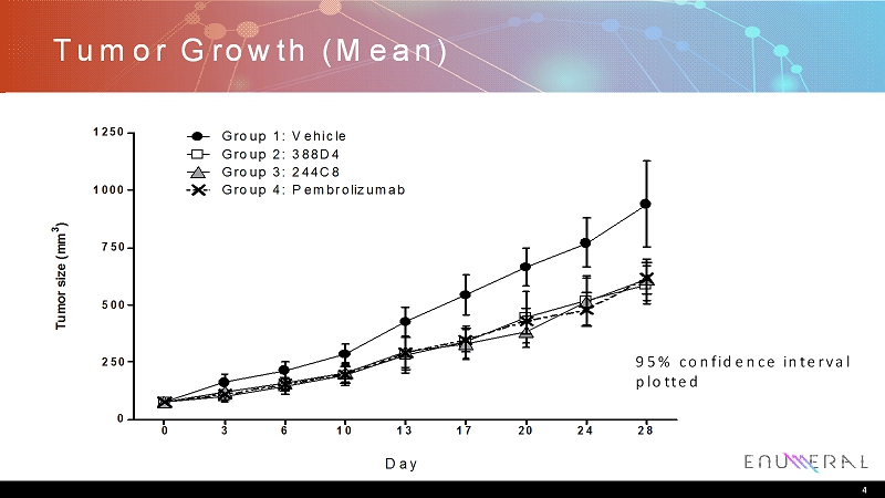
Tumor Growth (Mean) 4 Tumor size (mm 3 ) 0 3 6 10 13 17 20 24 28 0 250 500 750 1000 1250 Group 1: Vehicle Group 2: 388D4 Group 3: 244C8 Group 4: Pembrolizumab Day 95% confidence interval plotted

Study Observations • End of study tumor biopsies collected and analyzed • Low TIL % in tumors – <1% TIL in tumors – Suggests narrow dynamic range for immuno-modulation • Unclear if myeloid functionality in hu-NSG model – Kinetics of engraftment may not recapitulate biology of myeloid cells – Newer NSG-SGM3 model thought to rectify this 5

T Cell Infiltration from hu-NSG/PDX Tumors 6

Summary • Enumeralanti-PD1 antibodies 388D4 and 244C8 demonstrate activity in an accepted in vivo model of immunomodulation – huNSGlung PDxmodel used – Tumor growth inhibition similar to that observed with pembrolizumab • Initial characterization of tumor biopsy suggests low levels of TIL infiltration – Model has a narrow dynamic range for measuring activity of novel immunomodulatorsand may not reflect effects on non-T cell compartments 7

PD-1 Blockade in NSCLC Tumor Samples Complex IMR biology 8
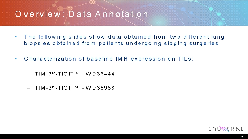
Overview: Data Annotation • The following slides show data obtained from two different lung biopsies obtained from patients undergoing staging surgeries • Characterization of baseline IMR expression on TILs: – TIM-3 lo /TIGIT lo -WD36444 – TIM-3 hi /TIGIT hi -WD36988 9

NSCLC WD36444 Contains 14% T Cells 10 CD45+ cells Live/dead cells T cells

WD36444 CD4 and CD8+ TILS Express PD1 hi , but TIM-3 lo CD4 cells CD8 cells P D - 1 TIM-3 • PD-1 is expressed on 55% of CD4 cells and 75% of CD8 cells • TIM-3 is expressed on 5.5% of CD4 cells and 13% of CD8 cells • Cells dually expressing both markers are observed 11

Ex Vivo PD-1 Blockade Can Drive Expression of TIM-3 on TILs Derived From Lung Cancer Biopsy Nivolumab P D - 1 TIM3 D4-2 C8-2 isotype stain control isotype stain control isotype stain control WD-36444 Condition CD3 + Tim-3% isotype <1 Nivolumab 31.4 D4-2 29.9 D4-3 34.7 C8-1 39.5 C8-2 27.3 C8-3 42.5 12

Ex Vivo PD-1 blockade Can Reverse TIL Exhaustion 13 Experimental Protocol: 300,000 cells purified from Tumor WD3644 were stimulated with anti-CD3+anti- CD28 in the presence of 20 ug/mL isotype control or the indicated anti-PD1 antibody. Supernatants were analyzed by ELISA after 24 hours of Activation. I s o t y p e C o n t r o l N i v o l u m a b E N U M D 4 - 2 E N U M D 4 - 3 E N U M C 8 - 1 E N U M C 8 - 2 E N U M C 8 - 3 0 10,000 20,000 30,000 40,000 I F N - ( p g / m L )

Selective IL-12 Induction with 244C8 in TIM-3 lo NSCLC N i v o l u m a b - 2 0 3 8 8 D 4 - 2 ( 2 0 u g / m L ) 3 8 8 D 4 - 3 ( 2 0 u g / m L ) 2 4 4 C 8 - 1 ( 2 0 u g / m L ) 2 4 4 C 8 - 2 ( 2 0 u g / m L ) 2 4 4 C 8 - 3 ( 2 0 u g / m L ) 0 500 1000 1500 WD-36444 IL-12 p70 p g / m l N i v o l u m a b - 2 0 3 8 8 D 4 - 2 ( 2 0 u g / m L ) 3 8 8 D 4 - 3 ( 2 0 u g / m L ) 2 4 4 C 8 - 1 ( 2 0 u g / m L ) 2 4 4 C 8 - 2 ( 2 0 u g / m L ) 0 10000 20000 30000 WD-36444 TNF- p g / m l 14
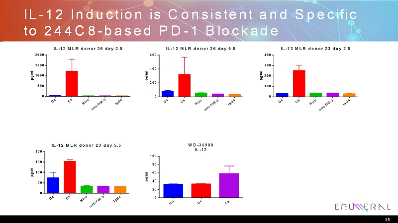
IL-12 Induction is Consistent and Specific to 244C8-based PD-1 Blockade D 4 C 8 N i v o a n t i - T I M - 3 I g G 4 0 500 1000 1500 2000 IL-12 MLR donor 26 day 2.5 p g / m l D 4 C 8 N i v o a n t i - T I M - 3 I g G 4 0 200 400 600 IL-12 MLR donor 26 day 5.5 p g / m l D 4 C 8 N i v o a n t i - T I M - 3 I g G 4 0 100 200 300 400 IL-12 MLR donor 23 day 2.5 p g / m l D 4 C 8 N i v o a n t i - T I M - 3 I g G 4 0 50 100 150 200 IL-12 MLR donor 23 day 5.5 p g / m l i s o D 4 C 8 0 20 40 60 80 100 WD-36988 IL-12 p g / m l 15
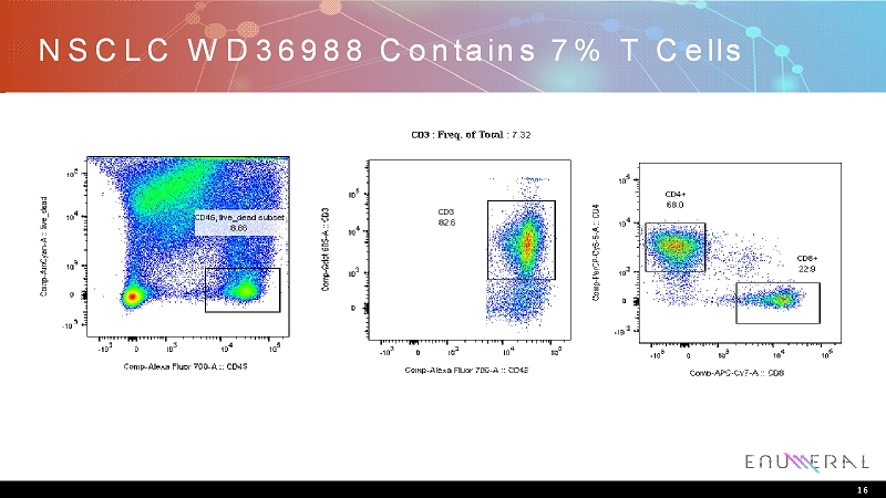
NSCLC WD36988 Contains 7% T Cells 16

CD8 + TILs Express PD-1,Tim-3,TIGIT 17 93% of CD8 + express PD-1 62% of CD8 + express TIM-3 No CD8 + express CEACAM-1 52 % of CD8 + express TIGIT 70 % of PD-1 + TIM3 + cells are also TIGIT + TIM-3 P D - 1 TIGIT TIGIT C E A C A M - 1 T i m - 3

Ex Vivo PD-1 Blockade can still increase IFNproduction in TIM-3 hi /TIGIT hi T cells B i o e g e n d I g G 4 ( 1 0 u g / m L ) 3 8 8 D 4 - 2 ( 1 0 u g / m L ) 2 4 4 C 8 - 2 ( 1 0 u g / m L ) 0 1000 2000 3000 4000 IFN- p g / m l B i o e g e n d I g G 4 ( 1 0 u g / m L ) 3 8 8 D 4 - 2 ( 1 0 u g / m L ) 2 4 4 C 8 - 2 ( 1 0 u g / m L ) 0 500 1000 1500 2000 Lung IL-2 p g / m l 18
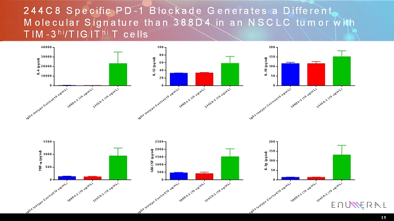
244C8 Specific PD-1 Blockade Generates a Different Molecular Signature than 388D4 in an NSCLC tumor with TIM-3 hi /TIGIT hi T cells I g G 4 I s o t y p e C o n t r o l ( 1 0 u g / m L ) 3 8 8 D 4 - 2 ( 1 0 u g / m L ) 2 4 4 C 8 - 2 ( 1 0 u g / m L ) 0 10000 20000 30000 40000 I L - 6 ( p g / m l ) I g G 4 I s o t y p e C o n t r o l ( 1 0 u g / m L ) 3 8 8 D 4 - 2 ( 1 0 u g / m L ) 2 4 4 C 8 - 2 ( 1 0 u g / m L ) 0 20 40 60 80 100 I L - 1 2 ( p g / m l ) I g G 4 I s o t y p e C o n t r o l ( 1 0 u g / m L ) 3 8 8 D 4 - 2 ( 1 0 u g / m L ) 2 4 4 C 8 - 2 ( 1 0 u g / m L ) 0 50 100 150 200 I L - 1 8 ( p g / m l ) I g G 4 I s o t y p e C o n t r o l ( 1 0 u g / m L ) 3 8 8 D 4 - 2 ( 1 0 u g / m L ) 2 4 4 C 8 - 2 ( 1 0 u g / m L ) 0 500 1000 1500 T N F - ( p g / m l ) I g G 4 I s o t y p e C o n t r o l ( 1 0 u g / m L ) 3 8 8 D 4 - 2 ( 1 0 u g / m L ) 2 4 4 C 8 - 2 ( 1 0 u g / m L ) 0 500 1000 1500 2000 2500 G M - C S F ( p g / m l ) I g G 4 I s o t y p e C o n t r o l ( 1 0 u g / m L ) 3 8 8 D 4 - 2 ( 1 0 u g / m L ) 2 4 4 C 8 - 2 ( 1 0 u g / m L ) 0 50 100 150 200 I L - 1 ( p g / m l ) 19

Summary • NSCLC tumors generally have PD-1 hi T cell infiltrates, however the level of additional checkpoint markers varies considerably • PD-1 biology appears to be linked to TIM-3 receptor expression and both markers appear to be important in T cell exhaustion in NSCLC tumors • PD-1 blockade can be biologically distinct between anti-PD-1 antibodies – Enumeral244C8 is an anti-PD-1 antibody that may promote release of non T cell (myeloid) derived anti-tumor cytokines 20

THE POWER ofHUMAN™