Attached files
| file | filename |
|---|---|
| 8-K - BIOMIMETIC THERAPEUTICS, INC. | v176836_8k.htm |
| EX-99.1 - BIOMIMETIC THERAPEUTICS, INC. | v176836_ex99-1.htm |
| EX-99.4 - BIOMIMETIC THERAPEUTICS, INC. | v176836_ex99-4.htm |
| EX-99.3 - BIOMIMETIC THERAPEUTICS, INC. | v176836_ex99-3.htm |

Evaluation of rhPDGF-BB in Combination with
a Flowable Collagen Matrix for the Treatment
of Acute Achilles Tendon Injury
1Hee, C K; 1Roden, C M; 1Wisner-Lynch, L A; 1Aguiar, D J; 2Dines, J S; 3Turner, A S;
3Ruehlman, D L; 1Kestler, H K; 1Lynch, S E; 3McGilvray, K C; 3Lyons, A S;
3Puttlitz, C M; 3Santoni, B G
1BioMimetic Therapeutics, Franklin, TN; 2Hospital for
Special Surgery, New York, NY;
3Colorado State University, Fort Collins, CO
chee@biomimetics.com

Disclosures
This study was funded by BioMimetic Therapeutics

Rerupture rates are high for the current repair treatments
Clinical Need for Improved Achilles Tendon Repair
(Leppilahti+, 1998; Maffulli, 1999)
Surgical Intervention
Conservative Treatment
Comparative Function
Equal or Increased
Equal or Decreased
Rerupture Rate
1 – 9%
8 – 39%
Prolonged immobilization has negative effects on the rerupture rate
Current methods require varying periods of immobilization
Shortening the immobilization time leads to better functional results but can increase the
rerupture rate

Injury
Rupture
Laceration
Phases of Tendon Repair
Sharma and Mafulli, 2005; James+, 2008
Inflammatory Phase (0d-1wk)
Clot formation releases chemotactic factors
Migration of macrophages, phagocytes,
fibroblasts
Fibroblastic/Proliferative Phase
(1wk-6wk)
Recruitment and proliferation of fibroblasts
Release of growth factors
Deposition of ECM
Remodeling Phase (6 wk +)
Deposition of ECM
Reorganization of collagen fibers
Increased collagen crosslinking

Growth Factors Stimulate Early Tendon Wound Healing
Hollinger+, 2008; Foster+, 2009
Injury
Platelets
TGF-ß
PDGF
FGF
EGF
VEGF
IGF
a-granules
Tendon Repair
Chemotaxis
Macrophages
Phagocytes
Fibroblasts
Mitogenesis
Fibroblasts
Angiogenesis
Vascular Smooth
Muscle Cells

Role of PDGF-BB in Tendon Repair
PDGF-BB is endogenously expressed in healing flexor tendons
(Ahlen+,1994; Brogi+,1994; Letson and Dahners,1994; Duffy+,1995; Banes+,1995; Batten+, 1996; Harwood+,1999;
Hildebrand+,1998; Yoshikawa+,2001; Affleck+,2002; Bouletreau+,2002; Tsubone+,2004; Weiler+,2004; Thomopoulos+,2005;
Chan+,2006; Costa+,2006; Thomopoulos+,2007, Sakiyama-Elbert+,2008; Thomopoulos+,2009; Thomopoulos+,
2010)
Repair Phase
Effect of Exogenous PDGF-BB
Inflammatory
av ß3, a5ß1, a2 ß1 integrins
VEGF
Fibroblastic/
Proliferative
DNA
Type I Collagen
ECM Synthesis
Collagen Crosslinks
PDGF-BB enhances function of injured ligaments and tendons in vivo

rhPDGF-BB combined with a collagen matrix in an acute ovine
Achilles tendon injury will:
Increase the biomechanical properties (load at failure, stiffness,
energy to failure) of the repaired tendon over the collagen matrix
alone
Improve the histological appearance (integration, collagen fiber
alignment, collagen fiber density) of the repaired tendon over the
collagen matrix alone
Hypothesis

Surgical Procedure
Acute Achilles transection in skeletally mature ewes
Single suture with a modified Mason-Allen knot in transected tendon ends
Lower leg was casted for 2 wks
8 wk survival time (n=8/group)
Group
Collagen
(5% w/v)
rhPDGF-BB
(µg)
Volume
(cc)
1
Yes
0
0.5
2
Yes
150
0.5
3
Yes
500
0.5
(Virchenko+, 2008; Sarrafian+, 2010)
Biomechanical Analysis (n=6)
Cyclic preconditioning (60 cycles, 0.25Hz, 10N-50N) followed by ramp-to-failure (1 mm/sec)
Load-to-failure (N), Repair Tissue Stiffness (N/mm), and Energy-to-Failure (J)
Histology (n=2)
H&E stained samples were evaluated to assess repair quality:
Native tendon/reparative tissue interface, quality of the reparative/healing tissue, collagen
density/fiber orientation
Statistics (for biomechanical properties)
One-way ANOVA with a post-hoc Tukey’s test (significance set at p<0.05)
Data are shown as Mean ± SEM
Analyses

Ultimate Force of the Repaired Tendon
p=0.082
57%
n=6
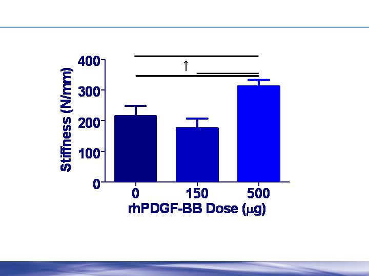
Repaired Tendon Stiffness
p=0.012
p=0.075
p=0.013
45%
n=6
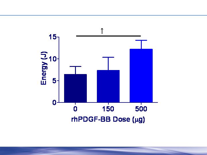
Energy to Failure of the Repaired Tendon
p=0.209
90%
n=6

Good Alignment and Integration of Tendon Repair
0 µg rhPDGF-BB
150 µg rhPDGF-BB
500 µg rhPDGF-BB
Dotted line shows site of transection
500 µm
Dose-dependent increases were observed for the biomechanical properties
Collagen+150 µg rhPDGF-BB was similar to matrix alone
Collagen+500 µg rhPDGF-BB compared to matrix alone
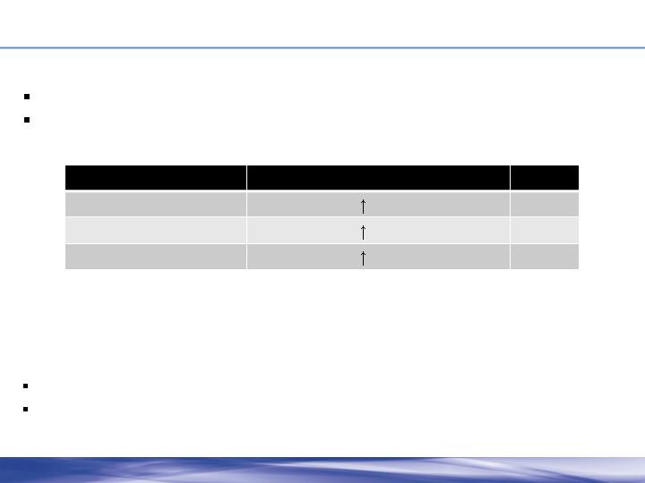
Discussion
Biomechanical Property
500 µg compared to matrix alone
P-value
Ultimate Force
57%
0.08
Stiffness
45%
0.08
Energy to Failure
90%
0.21
This is consistent with other studies showing the role of PDGF-BB in healing tendon and
ligament function (Letson and Dahners, 1994; Batten+, 1996;
Hildebrand+, 1998; Weiler+, 2004;
Chan+, 2006; Thomopoulos+, 2009)
Dose-dependent effects
Improvement of tendon function and biomechanics

Conclusion
The results of this study suggest that rhPDGF-BB
delivered in a collagen matrix has promise for the
treatment of Achilles tendon injuries

Acknowledgements